Research Snappy
  • Market Research Forum
  • Investment Research
  • Consumer Research
  • More
    • Advertising Research
    • Healthcare Research
    • Data Analysis
    • Top Companies
    • Latest News
No Result
View All Result
Research Snappy
No Result
View All Result

Patients with myeloproliferative neoplasms and high levels of systemic inflammation develop age-related macular degeneration

researchsnappy by researchsnappy
September 8, 2020
in Healthcare Research
0
Patients with myeloproliferative neoplasms and high levels of systemic inflammation develop age-related macular degeneration
400
SHARES
2.4k
VIEWS
Share on FacebookShare on Twitter

Abstract

Background

Epidemiological data show that myeloproliferative neoplasms (MPNs) are associated with increased risk of neovascular age-related macular degeneration (AMD). However, knowledge about the retinal findings in these patients is lacking. This study was conducted to examine retinal ageing and the prevalence of a hallmark of AMD; drusen, in patients with MPNs. Further, we examine the role of chronic systemic inflammation, considered central in both AMD and MPNs.

Methods

In this single-centre cross-sectional study, we consecutively enrolled 200 patients with MPNs. The study was divided into three substudies. Firstly, we obtained colour fundus photographs from all patients to evaluate and compare the prevalence of drusen with the published estimates from three large population-based studies. Secondly, to evaluate age-related changes in the various retinal layers, optical coherence tomography images were obtained from 150 of the patients and compared to a healthy control group, from a previous study. Thirdly, venous blood was sampled from 63 patients to determine the JAK2V617F allele burden and neutrophil-to-lymphocyte ratio (NLR), a marker of systemic inflammation, in MPN patients with and without drusen.

Findings

Patients with MPNs had an increased risk of having large drusen compared to the three population-based studies OR 5·7 (95%CI, 4·1–8·0), OR 6·0 (95%CI, 4·2–8·4) and OR 7·0 (95%CI, 5·0–9·7). Also, we found that the retinal site of drusen accumulation – the Bruch’s-membrane-retinal-pigment-epithelium-complex was thicker compared to healthy controls, 0·43μm (95%CI 0·17–0·71, p = 0·0014), but there was no sign of accelerated retinal ageing in terms of thinning of the neuroretina. Further, we found that MPN patients with drusen had a higher level of systemic inflammation than MPN patients with no drusen (p = 0·0383).

Interpretation

Patients with MPNs suffer from accelerated accumulation of subretinal drusen and therefore AMD from an earlier age than healthy individuals. We find that the retinal changes are located only between the neuroretina and the choroidal bloodstream. Further, we find that the drusen accumulation is associated with a higher JAK2V617F allele burden and a higher NLR, suggesting that low-grade chronic inflammation is a part of the pathogenesis of drusen formation and AMD.

Funding

Fight for Sight, Denmark and Region Zealand’s research promotion fund.

1. Introduction

The Philadelphia-negative myeloproliferative neoplasms (MPNs) are acquired clonal stem cell neoplasms and include essential thrombocythaemia (ET), polycythaemia vera (PV), and primary myelofibrosis (PMF). These diseases are characterised by an excess production of one or more of the mature blood cells from the myeloid lineage, and they cause massive systemic inflammation as well as neoangiogenesis and fibrosis in the bone marrow [

1

  • Hasselbalch H.C.
  • Bjørn M.E.
MPNs as inflammatory diseases: the evidence, consequences, and perspectives.

,

2

Inflammation and myeloproliferative neoplasms.

,

3

Myeloproliferative Neoplasms.

]. Recently it has been shown that patients with MPNs are at increased risk of neovascular age-related macular degeneration (AMD) [

4

  • Bak M.
  • Sørensen T.L.
  • Flachs E.M.
  • et al.
Age-related macular degeneration in patients with chronic myeloproliferative neoplasms.

], a progressive degenerative disease of the retina, causing loss of central vision. Worldwide AMD accounts for 8.7% of legal blindness, and it is the most common cause of visual impairment in the western world [

5

  • Wong W.L.
  • Su X.
  • Li X.
  • et al.
Global prevalence of age-related macular degeneration and disease burden projection for 2020 and 2040: a systematic review and meta-analysis.

]. The hallmark sign of AMD is drusen. Drusen consist of cellular debris located between the retinal pigment epithelium (RPE) and Bruch’s membrane. AMD-associated lesions: drusen and pigmentary abnormalities, characterise the early stages of AMD. In addition, the late stages of AMD have either characteristic neovascular lesions or sharply demarcated areas of retinal atrophy, defined as neovascular AMD and geographic atrophy (GA), respectively [

6

  • Cook H.L.
  • Patel P.J.
  • Tufail A
Age-related macular degeneration: diagnosis and management.

]. Despite extensive research, the exact pathophysiology of AMD remains unknown. Certain risk factors are known such as age, cigarette smoking and genetic susceptibility [

7

  • Tomany S.C.
  • Wang J.J.
  • Van Leeuwen R.
  • et al.
Risk factors for incident age-related macular degeneration: pooled findings from 3 continents.

,

8

  • Smith W.
  • Assink J.
  • Klein R.
  • et al.
Risk factors for age-related macular degeneration: pooled findings from three continents.

]. In recent years evidence have emerged demonstrating the role of the immune system, including inflammation, in the pathogenesis of the disease [

9

  • Kauppinen A.
  • Paterno J.J.
  • Blasiak J.
  • Salminen A.
  • Kaarniranta K
Inflammation and its role in age-related macular degeneration.

,

10

  • Ambati J.
  • Atkinson J.P.
  • Gelfand B.D
Immunology of age-related macular degeneration.

,

11

  • Chen M.
  • Parainflammation Xu H.
chronic inflammation, and age-related macular degeneration.

,

12

Aging is not a disease: distinguishing age-related macular degeneration from aging.

,

13

  • Fernandez-Robredo P.
  • Recalde S.
  • Hernandez M.
  • et al.
Novel association of high C-reactive protein levels and A69S at risk alleles in wet age-related macular degeneration women.

,

14

  • Nita M.
  • Grzybowski A.
  • Ascaso F.J.
  • Huerva V
Age-related macular degeneration in the aspect of chronic low-grade inflammation (pathophysiological parainflammation).

,

15

  • Parmeggiani F.
  • Sorrentino F.S.
  • Romano M.R.
  • et al.
Mechanism of inflammation in age-related macular degeneration: an up-to-date on genetic landmarks.

,

16

Inflammation and age-related macular degeneration (AMD).

,

17

Systemic inflammatory disease and AMD comorbidity.

, ]. Since MPNs cause massive inflammation and a recent register study has reported that patients with MPNs have a significantly higher risk of neovascular AMD [

4

  • Bak M.
  • Sørensen T.L.
  • Flachs E.M.
  • et al.
Age-related macular degeneration in patients with chronic myeloproliferative neoplasms.

], it is intriguing to investigate these patients in the context of AMD.

The two disease groups, MPNs and AMD, have important similarities such as inflammation, angiogenesis, and fibrosis. However, knowledge of the retinal status in patients with MPNs is lacking. We are unaware if MPNs are merely associated with angiogenesis, or if the occurrence of other AMD-associated lesions is also increased in these patients. With this study, we investigated an unselected cohort of patients with MPNs. As the first substudy, we evaluated fundus photographs to assess AMD-associated lesions and thereby the prevalence of different AMD-stages. In substudy two, we further investigated the retinas of these patients with optical coherence tomography (OCT) images to evaluate the age-related changes in the structural layers. In substudy three, we examined the role of chronic inflammation in patients with MPNs, through measurement of the neutrophil-to-lymphocyte ratio (NLR), a marker of systemic inflammation, previously shown to be elevated in patients with AMD [

19

  • Niazi S.
  • Krogh Nielsen M.
  • Sørensen T.L.
  • Subhi Y
Neutrophil-to-lymphocyte ratio in age-related macular degeneration: a systematic review and meta-analysis.

]. Further, we assessed the JAK2V617F allele burden as a marker of inflammation, since the JAK2V617F mutation is seen as a key driver of MPN-associated chronic inflammation [

20

  • Lussana F.
  • Carobbio A.
  • Salmoiraghi S.
  • et al.
Driver mutations (JAK2V617F, MPLW515L/K or CALR), pentraxin-3 and C-reactive protein in essential thrombocythemia and polycythemia vera.

].

2. Methods

2.1 Study design and participants

This cross-sectional study was approved by the Ethics Committee in Region Zealand, Denmark (SJ-588, SJ-679), and each patient provided oral and written informed consent. The eligibility criterion was a diagnosis of a Philadelphia-negative MPN (WHO2016 criteria) [

21

  • Barbui T.
  • Thiele J.
  • Gisslinger H.
  • et al.
The 2016 WHO classification and diagnostic criteria for myeloproliferative neoplasms: document summary and in-depth discussion.

]. Patients at the Department of Haematology, Zealand University Hospital (ZUH), were invited to participate in the study. Patients were consecutively enrolled for substudy one until we reached 200, and inclusion was conducted between January 2017 and October 2019. Of the 200 patients, 150 were enrolled in substudy two to match the number in the healthy control group. Finally, 63 of the patients not receiving immunomodulating treatment were enrolled in substudy three.

In substudy one, drusen prevalence data were compared to the published estimates from three population-based studies; the Beaver Dam Eye Study (BDES), The Blue Mountains Eye Study (BMES) and The Rotterdam Study (RS) [

22

  • Mitchell P.
  • Smith W.
  • Attebo K.
  • Wang J.J
Prevalence of Age-related Maculopathy in Australia: the Blue Mountains Eye Study.

,

23

  • Klein R.
  • Klein B.E.
  • Linton K.L
Prevalence of age-related maculopathy. The Beaver Dam Eye Study.

,

24

  • Vingerling J.R.
  • Dielemans I.
  • Hofman A.
  • et al.
The prevalence of age-related maculopathy in the rotterdam study.

].

In substudy two, we compared layers in the retina with a healthy aged control group of 150 patients. The control group was from a previous study by Harris et al., and characteristics and inclusion criteria are described thoroughly elsewhere [

25

  • Harris J.
  • Subhi Y.
  • Sørensen T.L
Effect of aging and lifestyle on photoreceptors and retinal pigment epithelium: cross-sectional study in a healthy Danish population.

]. This control group did not have fundus photographs taken and could not be used as controls for substudy one.

In substudy three NLR and the key driver of MPN-associated chronic inflammation JAK2V617F mutation was compared between MPN patients with drusen and those without.

2.2 Imaging and grading method

All participants underwent an examination at the Department of Ophthalmology, ZUH and had their pupils dilated with tropicamide 1% before examination. For substudy one, we obtained a stereoscopic 45° colour fundus photograph centred on the macula (model TRG-NW8, Topcon). Each fundus photograph was graded using a simplified version of the Wisconsin age-related maculopathy grading system (WARMGS) [

26

  • Klein R.
  • Davis M.D.
  • Magli Y.L.
  • Segal P.
  • Klein B.E.
  • Hubbard L
The Wisconsin age-related maculopathy grading system.

] (supplementary material 1.1). This allows comparisons to several studies, including the population-based studies used in this study. We chose to compare our results to populations of European ancestry since ethnicity differences have been reported [

5

  • Wong W.L.
  • Su X.
  • Li X.
  • et al.
Global prevalence of age-related macular degeneration and disease burden projection for 2020 and 2040: a systematic review and meta-analysis.

].

We applied the classification system introduced by the Beckman Initiative for Macular Research Classification Committee [

27

  • Ferris F.L.
  • Wilkinson C.P.
  • Bird A.
  • et al.
Clinical classification of age-related macular degeneration.

] using drusen size and presence of pigmentary abnormalities to classify AMD (supplementary material 1.3).

One investigator (CL) graded all images, and another investigator (MKN) re-graded 80 images to test intergrader agreement.

The types of drusen present were categorised in three groups according to the description from Spaide and Curcio; soft drusen, cuticular drusen and subretinal drusenoid deposits [

28

Drusen characterization with multimodal imaging.

] (supplementary material 1.1).

For substudy two, we obtained optical coherence tomography images (SD-OCT, Heidelberg Engineering, Heidelberg, Germany) from 150 of the patients. The images were examined in Heidelberg Eye Explorer version 1.9.10.0. We used the automated segmentation and the thickness profile part of the software to measure the thickness of the different retinal layers, and segmentations was checked manually (supplementary material 1.2).

The neuroretina is the inner part of the retina and can be subdivided into several layers. The retinal pigment epithelium (RPE) is a monolayer of pigmented cells in the outer retina, essential for homoeostasis of the retina and vision. The Bruch’s membrane (BM) is the innermost layer of the choroid were the fenestrated capillaries of the eyes are located. The inner part of the BM forms the basement membrane of the RPE (supplementary material 1.2). The thickness of the neuroretina and RPE-BM-complex were compared to values measured from a healthy aged control group of 150 patients [

25

  • Harris J.
  • Subhi Y.
  • Sørensen T.L
Effect of aging and lifestyle on photoreceptors and retinal pigment epithelium: cross-sectional study in a healthy Danish population.

].

2.3 Blood sampling

For substudy three, venous blood from antecubital veins was sampled from 63 patients, not receiving immunomodulating treatment – 35 of the patients had drusen, and 28 had normal ageing changes. The blood was sampled in an ethylenediaminetetraacetic acid-coated (EDTA) tube and analysed on Sysmex KX-21NTM (Sysmex Corporation, Kobe, Japan), to measure white blood cells count; lymphocytes count, and percentage; neutrophils count, and percentage; and monocytes–basophils–eosinophils mixed count and percentage. Sample volume for the count was 50 µl. NLR was calculated by dividing the absolute neutrophil count by the absolute lymphocyte count. The JAK2V617F mutation analysis was performed on peripheral blood EDTA anticoagulated blood with highly sensitive real-time quantitative PCR on an ABI Prism7900HT (Applied Biosystems, Foster City, CA, USA), on fluorescence-activated cell sorted (FACS) monocytes, lymphocytes and granulocytes on a FACSVantage (BD Biosciences). DNA was extracted using a MagnaPure robot (Roche Diagnostics, Mannheim, Germany) according to the manufacturer’s protocol.

2.4 Outcomes

The main outcomes were drusen size (largest present), pigmentary abnormalities, early-, intermediate- and late AMD, neuroretinal- and RPE-BM thickness, NLR and JAK2V617F allele burden. Secondary outcomes were area-covered-by drusen; drusen count, and drusen type.

2.5 Statistical analysis

To detect a difference of at least 20%, at a 5% significance level and a power of 80%, the total sample size required is 140–210 []. We aimed for 200 in substudy one and 150 in substudy two. Power calculations for substudy three were based upon previous immunologic studies of neovascular AMD were calculations show a sample size of minimum 26 in each group is necessary [

30

  • Krogh Nielsen M.
  • Subhi Y.
  • Molbech C.R.
  • et al.
Patients with a fast progression profile in geographic atrophy have increased CD200 expression on circulating monocytes.

].

We analysed the data using the SAS statistical software package (SAS ver. 9.4; SAS Institute Inc.).

In substudy one, the age-associated prevalence rates were compared with the estimates from the population studies by using the Chi-squared test. To assess interobserver-agreement in image-grading, we calculated Cohen’s kappa coefficient [

31

Interrater reliability: the kappa statistic.

]. We found strong agreement for drusen size (weighted kappa=0·87) and drusen count (weighted kappa=0·86), moderate agreement for drusen area (weighted kappa=0·79) and weak agreement for pigment abnormalities (weighted kappa=0·47).

In substudy two and three, linear regression models with age as exposure variable were used to investigate the relationship between the continuous outcome variables; layer thicknesses of the retina, NLR and JAK2V617F allele burden. Non-normal data were transformed. Where outcome was independent of age, a two-sided t-test was used for normally distributed data and Wilcoxon rank-sum test for non-normal data. Otherwise, analysis of covariance (ANCOVA) was used to estimate differences between the two regression lines corresponding to groups.

2.6 Role of the funding source

The funding sources had no role in the design and conduct of the study; collection, analysis, and interpretation of the data; preparation, review, or approval of the manuscript; or the decision to submit the manuscript for publication. The corresponding author confirms that she had full access to all the data in the study and had final responsibility for the decision to submit for publication.

3. Results

3.1 Study population

For substudy one, 200 patients were enrolled. One was excluded post hoc since the diagnosis of MPN was later questioned. Eight patients were excluded due to poor image quality of the obtained fundus photographs. As a result, 191 patients had gradable photographs and were included for analyses.

For substudy two, we obtained OCT images for 150 of the patients. Seven patients with late AMD and four with poor image quality were excluded. As a result, 139 were included for further analyses. The healthy control group used in substudy two had no ocular or systemic diseases known to influence the macula (cancer, blood diseases, severe hypertension or diabetes) and they had normal macular ageing changes (no or few drusen). For more information on the control group see Harris et al. [

25

  • Harris J.
  • Subhi Y.
  • Sørensen T.L
Effect of aging and lifestyle on photoreceptors and retinal pigment epithelium: cross-sectional study in a healthy Danish population.

]

For substudy three, we collected blood samples from 63 of the patients.

Patient characteristics are shown in Table 1. The majority of the patients suffered from PV and to a lesser extent ET and PMF. Most patients had the JAK2V617F mutation, associated with MPNs. The patients received different treatments for their MPNs; Peginterferon alfa-2a, Ruxolitinib or a combination, hydroxyurea, anagrelide, acetylsalicylic acid or other drugs with anticoagulant effect, and statins. It was not possible for us to assess the potential effect of treatment on retinal changes since detailed information regarding treatment, was not available. We did not have information on previous treatment or for how long the treatments had been administered.

Table 1Characteristics of the participants in substudy 1, 2 and 3.

PV: Polycythaemia vera, ET: Essential thrombocythemia, PreMF: Pre-myelofibrosis, PMF: primary myelofibrosis JAK2V617F: mutation in the JAK2 gene, CALR: calreticulin gene, MPL: MPL gene, gene encoding the thrombopoietin receptor.

3.2 Substudy 1 – AMD-associated lesions and AMD-stages

Drusen size (largest present): For MPN patients younger than 54 years, the most frequent finding was small drusen less than 63μm. With increasing age, the prevalence of larger drusen increased, and the prevalence of small drusen correspondingly decreased. Large drusen greater than 125μm increased from 10% in patients younger than 45 years to 45% in patients 75–84 years (Fig. 1).

Fig. 1

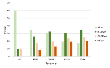
Fig. 1Age-specific distribution of maximum drusen size within a radius of 3000 µm from the fovea of the worst eye in patients with myeloproliferative neoplasms.

Drusen count: Only 6·4% of patients with MPNs had no drusen in the macular area, 34·2% had between one to nine drusen, and 59·4% had ten or more drusen. The number of drusen increased with increasing age. Ten or more drusen were seen in 20·0% of patients younger than 45 years and 71·8% of the patients aged 75–84 years.

Drusen type: For patients with drusen larger than 63 μm (129 patients), the drusen-type were primarily soft drusen (91·1%). In 11 patients, both soft drusen and one of the two types, cuticular drusen or subretinal drusenoid deposits, were also found.

Drusen area: The area-covered-by drusen within the grading-grid was greater than 0·069 mm2 in 49·7% of the patients, greater than 0·146 mm2 for 26·5%, greater than 0·487 mm2 for 12·4%, greater than 1·27 mm2 for 4·9% and greater than 2·5 mm2 for 2·2% of the patients.

Pigmentary abnormalities: Of the 184 patients without late AMD, we found pigmentary abnormalities in 25 cases (13·6%; CI 9·4−19·3%). Nine had increased pigment, nine had hypopigmentation, and seven had combined types. Accordingly, 16 patients (8·7%) had increased pigment, and 16 patients (8·7%) had decreased pigment. None of the patients younger than 45 years had pigmentary abnormalities. For the age groups 45–54, 55–64, 65–74 and 75–84 years, the prevalence was 13·6%, 4·3%, 17·1% and 22·2%, respectively. Except for the age group 55–64 years, pigment abnormalities were increasing with age.

Presence of AMD: Table 2 shows the prevalence of AMD stages. The prevalence of intermediate and late AMD increased with age. The prevalence of early AMD increased with age in patients up to 55–64 years. For patients older than 65 years, the prevalence of early AMD decreases as the prevalence of intermediate and late AMD increases. Late AMD was seen in seven patients (3·7%; CI, 1·8%−7·4%), six had geographic atrophy, and one had neovascular AMD. In patients younger than 45 years and patients aged 55–64 years, none had late AMD. For the age groups 45–54 years, 65–74 years and 75–84 years, the prevalence was 4·4%, 2·8% and 10·0%, respectively.

Table 2Age-specific prevalence of all stages of age-related macular degeneration (AMD) in patients with MPNs and the prevalence rates of late AMD in the same age groups from The Beaver Dam Eye Study, The Blue Mountains Eye Study and the Rotterdam Eye Study.

MPN: myeloproliferative neoplasms.

The age groups in The Beaver Dam Eye Study and The Blue Mountains Eye study are different from the age groups in this study: 1age group 43–54 years. 2age group 75–86 years. 3age group 49–54 years. 4The Rotterdam Study did not include patients younger than 55 years.

Since the classification of the earlier stages of AMD is different in our study compared to the population-based studies, it is not possible to compare these stages.

3.3 Results compared to prevalence rates from population studies

Drusen size: Fig. 2 compares the prevalence of large drusen (>125 µm) between patients with MPNs and the population studies. The figure further shows that patients with MPNs at the age of 45–54 years have the same prevalence of large drusen as a population that is 75–84 years old, indicating that patients with MPNs develop intermediate AMD at an earlier age. The prevalence rates of large drusen were significantly higher in patients with MPNs (p-valuesTable 3.

Fig. 2

Fig. 2Comparison of the prevalence of drusen >125 μm as the largest drusen present within a 3000 μm radius of the fovea between patients with Myeloproliferative neoplasms and three large population-based studies (Beaver Dam Eye Study, Blue Mountains Eye Study and Rotterdam Study).

Table 3Age-specific odds ratios for large drusen >125 µm within a 3000 µm radius of the fovea for MPN patients compared to the Beaver Dam Eye Study, The Blue Mountains Eye Study and The Rotterdam Study.

Only RS gives the exact prevalence of drusen size 63–125 µm; 35·7%, 43·2%, 41·0% in the age groups 55–64 years, 65–74 years and 75–84 years, respectively. The comparable prevalence in MPN patients was 32·6%, 31·4% and 38·9%. There was no significant difference between MPN patients and the RS population in intermediate-sized drusen (P = 0.11).

Drusen area: The area-covered-by drusen within the grading-grid was greater than 0·069 mm2 in 49·7% of the patients and greater than 1·27 mm2 for 4·9% of the patients. These areas correspond to approximately to 0·3% and 4·5% of the macula area. In BMES similar numbers are given. Drusen covered more than 0·2% in 15·3% of the participants and more than 4·7% in 6·2% of the participants.

Pigmentary abnormalities: To compare with BDES and BMES, we excluded the age group younger than 45 years and patients with late AMD. Twenty-five (14·4%) of the MPN patients had hyperpigmentation, hypopigmentation or both. In the BDES and BMES, the prevalence of any pigmentary abnormality was 13·1% and 11·9% (data was not available to exclude patients aged 85+ in the BDES). There were no significant differences between the prevalence of any pigmentary abnormalities in patients with MPNs and the BDES (OR 1·1, CI 0·7–1·7) or the BMES (1·2, CI 0·8–1·9).

Excluding further the age-group 45–54 years to compare with the RS population, the prevalence of any pigmentary abnormalities in patients with MPNs was 14·5%. The prevalence in the RS study was 7·0% (age group 85+ excluded). Patients with MPNs had a significantly higher risk of pigmentary abnormalities compared to the RS population (OR 2·2, CI 1·4–3·5).

In patients with MPNs, 8·7% had hyperpigmentation, and 8·7% had hypopigmentation. These numbers for hyper- and hypopigmentation was for BDES 12·2% and 8·3%, BMES 12·1% and 5·8%, RS 5·9% and 4·4%.

Patients with MPNs had a significantly higher risk of having hypopigmentation compared to the BMES (OR 1·8, CI 1·0–3·0) and a significantly higher risk of both hyper- and hypopigmentation compared to the RS population (OR 1·8, CI 1·1–3·1) (OR 2·0, CI 1·1–3·6).

Late AMD: Patients with MPNs had a significantly higher prevalence of late AMD compared to BDES, BMES and RS (Table 2) (p-values=0·0176, 0·0063, 0·0079). Patients with MPNs have a higher risk of late AMD compared to BDES (OR 2·5 [CI 1·1–5·5]), BMES (OR 2·9 [CI 1·3–6·6]) and RS (OR 2·9 [CI 1·3–6·8]). Only one patient with MPN had neovascular AMD, and six had geographic atrophy (GA), which is opposite to the population studies where neovascular AMD was found to have a higher prevalence than GA.

3.4 Substudy 2 – retinal thickness

The thicknesses of the retinal layers were approximately normally distributed. The RPE-BM layer was 0·43μm (ANCOVA, 95%CI 0·17–0·71, p = 0·0014) thicker in patients with MPNs compared to the healthy control group. Mean RPE-BM thickness in the macula for patients with MPNs was 14·39μm (95%CI 14·19–14·58) compared to 13·96μm (95%CI 13·75–14·16) for the healthy control group (which was older than the MPN patients, p-value <0·0001)

There was no significant difference in neuroretinal macular thickness between patients with MPNs and the healthy control group (ANCOVA, p-value=0·2990). Mean neuroretinal macular thickness in patients with MPNs was 286·55 μm (95%CI 283·85–289·25) compared to 288·95μm (95%CI 286·54–291·36) for the healthy controls.

We also evaluated each sublayer of the neuroretina in the centre-, inner and outer subfield independently (supplementary material 2).

3.5 Substudy 3 – chronic systemic inflammation and drusen formation

Neutrophil-to-lymphocyte ratio was not normally distributed and log-transformed. Linear regression lines were fitted for each group and showed that NLR was not dependent on age. We found that MPN patients with drusen had a significantly higher NLR than MPN patients without drusen, estimated difference 1·37 (T-test, 95%CI 1·02–1·86, p = 0·0383).

The JAK2V617F allele burden was non-normally distributed and did not fit an approximately normal distribution with transformation. Linear regression showed that the allele burden was not dependent on age. We found a higher allele burden of JAK2V617F among patients with MPNs and drusen (Wilcoxon rank-sum test, P = 0·0754). Thirty-three out of 35 patients (94·3%) with MPNs and drusen had the JAK2V617F mutation, and the median allele burden was 33·00 [IQR 11·00–56·00]. Only 24 out of 28 of the patients (85·7%) without drusen had the JAK2V617F mutation, and the median allele burden was 17·50 [IQR 5·55–28·50].

4. Discussion

In this study of patients with Philadelphia-negative MPNs, we found a higher prevalence of large drusen and AMD compared to estimates from large population-based studies. In addition, the retinal changes appear at an earlier age in patients with MPNs. Our data support the findings in a register study by Bak et Al [

4

  • Bak M.
  • Sørensen T.L.
  • Flachs E.M.
  • et al.
Age-related macular degeneration in patients with chronic myeloproliferative neoplasms.

], showing that MPN patients are at increased risk of having neovascular AMD. Further, we find that MPNs are not only related to neovascularization but also to the subretinal accumulation and the formation of drusen. We found no difference in the neuroretina suggesting that the retinal changes are not due to overall accelerated ageing of the retina, but rather to the accumulation of debris between the neuroretina and the choroidal bloodstream.

Studies of the natural history have shown that the risk of developing late AMD increases with advancing age and that the number and size of drusen also possess predictive value. Drusen volume, bilateral drusen and pigment abnormalities further increase the risk of late AMD [

32

  • Chew E.Y.
  • Clemons T.E.
  • Agrón E.
  • et al.
Ten-year follow-up of age-related macular degeneration in the age-related eye disease study: AREDS report No. 36.

,

33

  • Abdelfattah N.S.
  • Zhang H.
  • Boyer D.S.
  • et al.
Drusen volume as a predictor of disease progression in patients with late age-related macular degeneration in the fellow eye.

]. Patients with MPNs have large drusen in their macular area, and a higher prevalence of late AMD compared to population studies. In our study, we found a higher occurrence of GA than neovascular AMD. The register study by Bak et al. reports an increased risk of neovascular AMD, but our results indicate that GA could be even more prevalent.

Numerous studies have shown that low-grade chronic inflammation is involved in the pathogenesis of AMD [

9

  • Kauppinen A.
  • Paterno J.J.
  • Blasiak J.
  • Salminen A.
  • Kaarniranta K
Inflammation and its role in age-related macular degeneration.

,

10

  • Ambati J.
  • Atkinson J.P.
  • Gelfand B.D
Immunology of age-related macular degeneration.

,

11

  • Chen M.
  • Parainflammation Xu H.
chronic inflammation, and age-related macular degeneration.

,

34

  • Anderson D.H.
  • Mullins R.F.
  • Hageman G.S.
  • Johnson L V
A role for local inflammation in the formation of drusen in the aging eye.

,

35

  • Shankar A.
  • Mitchell P.
  • Rochtchina E.
  • Tan J.
  • Wang J.J
Association between circulating white blood cell count and long-term incidence of age-related macular degeneration: the Blue Mountains Eye Study.

,

36

Probing the role of inflammation in age-related macular degeneration.

,

37

  • Hageman G.S.
  • Luthert P.J.
  • Victor Chong N.H.
  • Johnson L V.
Anderson DH, Mullins RF. An integrated hypothesis that considers drusen as biomarkers of immune-mediated processes at the RPE-Bruch’s membrane interface in aging and age-related macular degeneration.

,

38

  • Klein R.
  • Myers C.E.
  • Cruickshanks K.J.
  • et al.
Markers of inflammation, oxidative stress, and endothelial dysfunction and the 20-year cumulative incidence of early age-related macular degeneration: the Beaver Dam Eye Study.

,

39

  • Park D.H.
  • Connor K.M.
  • Lambris J.D
The challenges and promise of complement therapeutics for ocular diseases.

] and chronic inflammation and immune deregulation are common features between AMD and MPNs [

1

  • Hasselbalch H.C.
  • Bjørn M.E.
MPNs as inflammatory diseases: the evidence, consequences, and perspectives.

,

9

  • Kauppinen A.
  • Paterno J.J.
  • Blasiak J.
  • Salminen A.
  • Kaarniranta K
Inflammation and its role in age-related macular degeneration.

,

45

  • Nussenblatt R.B.
  • Ferris F.
Age-related macular degeneration and the immune response: implications for therapy.

,

46

  • Jarrett S.G.
  • Boulton M.E.
Consequences of oxidative stress in age-related macular degeneration.

,

11

  • Chen M.
  • Parainflammation Xu H.
chronic inflammation, and age-related macular degeneration.

,

35

  • Shankar A.
  • Mitchell P.
  • Rochtchina E.
  • Tan J.
  • Wang J.J
Association between circulating white blood cell count and long-term incidence of age-related macular degeneration: the Blue Mountains Eye Study.

,

38

  • Klein R.
  • Myers C.E.
  • Cruickshanks K.J.
  • et al.
Markers of inflammation, oxidative stress, and endothelial dysfunction and the 20-year cumulative incidence of early age-related macular degeneration: the Beaver Dam Eye Study.

,

40

  • Hermouet S.
  • Bigot-Corbel E.
  • Gardie B
Pathogenesis of myeloproliferative neoplasms: role and mechanisms of chronic inflammation.

,

41

  • Wang J.C.
  • Sindhu H.
  • Chen C.
  • et al.
Immune derangements in patients with myelofibrosis: the role of Treg, Th17, and sIL2Rα.

,

42

  • Anderson D.H.
  • Radeke M.J.
  • Gallo N.B.
  • et al.
The pivotal role of the complement system in aging and age-related macular degeneration: hypothesis re-visited.

,

43

  • Geyer H.L.
  • Dueck A.C.
  • Scherber R.M.
  • Mesa R.A
Impact of inflammation on myeloproliferative neoplasm symptom development.

,

44

  • Mitta V.P.
  • Christen W.G.
  • Glynn R.J.
  • et al.
C-reactive protein and the incidence of macular degeneration: pooled analysis of 5 cohorts.

]. Thus, chronic inflammation has been proposed as both a trigger and a driver of clonal evolution in MPNs [

47

Perspectives on the increased risk of second cancer in patients with essential thrombocythemia, polycythemia vera and myelofibrosis.

]. The neoplastic clone is a major source of inflammatory cytokines, released into the systemic circulation, and contributing to the symptom burden in patients with MPNs, and the inflammation-mediated comorbidities, including cerebral- and cardiovascular diseases, the increased risk of autoimmune diseases and second cancer [

47

Perspectives on the increased risk of second cancer in patients with essential thrombocythemia, polycythemia vera and myelofibrosis.

,

48

Perspectives on chronic inflammation in essential thrombocythemia, polycythemia vera, and myelofibrosis: is chronic inflammation a trigger and driver of clonal evolution and development of accelerated atherosclerosis and second cancer.

]. Similar to MPNs, AMD has been associated with systemic diseases characterised by immune modulation or inflammation, such as diabetes, cardiovascular disease and AIDS [

7

  • Tomany S.C.
  • Wang J.J.
  • Van Leeuwen R.
  • et al.
Risk factors for incident age-related macular degeneration: pooled findings from 3 continents.

,

49

  • Chen X.
  • Rong S.S.
  • Xu Q.
  • et al.
Diabetes mellitus and risk of age-related macular degeneration: a systematic review and meta-analysis.

,

50

Pennington KL, Deangelis MM. Epidemiology of age-related macular degeneration (AMD): associations with cardiovascular disease phenotypes and lipid factors. doi:10.1186/s40662-016-0063-5.

,

51

  • Jabs D.A.
  • Van Natta M.L.
  • Sezgin E.
  • Pak J.W.
  • Danis R
Prevalence of intermediate-stage age-related macular degeneration in patients with the acquired immunodeficiency syndrome HHS public access.

]. Rozing et al. have proposed a two-level hypothesis of the development of age-related degenerative diseases, including AMD []. The hypothesis includes two steps: the first step is an accumulation of retinal damage due to ageing, and the second step is the following inflammatory host response to these damages. Both steps should be present to develop AMD. In patients with MPNs, inflammation plays a pivotal role in disease pathogenesis and therefore possesses a massive “second step” contribution to the development of AMD. NLR is a good and relatively stable indicator of subclinical systemic inflammation [

52

  • Qin B.
  • Ma N.
  • Tang Q.
  • et al.
Neutrophil to lymphocyte ratio (NLR) and platelet to lymphocyte ratio (PLR) were useful markers in assessment of inflammatory response and disease activity in SLE patients.

], and we found that MPN patients with drusen have a significantly higher NLR and a tendency of higher JAK2-allele burden than MPN patients without drusen, further supporting a role for chronic inflammation in drusen formation and the pathogenesis of AMD.

In this context, it is intriguing to consider chronic inflammation as a major contributing common factor in disease pathogenesis between AMD and MPNs. Using the MPNs as a Human Inflammation Model [

1

  • Hasselbalch H.C.
  • Bjørn M.E.
MPNs as inflammatory diseases: the evidence, consequences, and perspectives.

,

53

  • Andersen M.
  • Sajid Z.
  • Pedersen R.K.
  • et al.
Mathematical modelling as a proof of concept for MPNs as a human inflammation model for cancer development.

] comparative molecular, genomic and immunological studies between drusen/AMD and MPNs are envisaged to unravel novel insights into disease-promoting mechanisms and how to modify disease evolution by targeting the common denominator for disease evolution and progression – chronic inflammation.

Another feature that occur during development of AMD is accumulation of mononuclear phagocytes in the subretinal space (between the photoreceptor’s outer segments and the RPE). The subretinal space is normally immune privileged and when mononuclear phagocytes occasionally reach the space in healthy individuals, these are eliminated by the immune-suppressive properties of the RPE. Animal models show that the mononuclear cells that infiltrate the subretinal space are likely resident microglia but also blood derived, monocyte derived inflammatory macrophages [

54

  • Guillonneau X.
  • Eandi C.M.
  • Paques M.
  • Sahel J.-.A.
  • Sapieha P.
  • Sennlaub F
On phagocytes and macular degeneration.

]. In donor eyes of patients with late AMD, the same mononuclear cells have been found to accumulate [

55

  • Gupta N.
  • Brown K.E.
  • Milam A.H
Activated microglia in human retinitis pigmentosa, late-onset retinal degeneration, and age-related macular degeneration.

].

Microglia respond to neuronal damage, and they increase in number and change morphology due to injury, ageing and disease. Microglia release inflammatory mediators and phagocytose and remove cellular debris [

56

  • Ellis-Behnke R.G.
  • Jonas R.A.
  • Jonas J.B
The microglial system in the eye and brain in response to stimuli in vivo.

,

57

  • Rashid K.
  • Wolf A.
  • Langmann T
Microglia activation and immunomodulatory therapies for retinal degenerations.

]. The components of drusen have been shown to retain microglia in the subretinal space and thereby sustain the inflammatory response which turns destructive and can influence photoreceptor and RPE integrity [

58

  • Ma W.
  • Coon S.
  • Zhao L.
  • Fariss R.N.
  • Wong W.T
A2E accumulation influences retinal microglial activation and complement regulation.

].

The chronic pro-inflammatory environment in patients with MPNs could also contribute to the overactivation of microglia by promoting blood-derived inflammatory mononuclear phagocytes with characteristics that lead to resisted elimination and excessive recruitment in the subretinal space [

54

  • Guillonneau X.
  • Eandi C.M.
  • Paques M.
  • Sahel J.-.A.
  • Sapieha P.
  • Sennlaub F
On phagocytes and macular degeneration.

].

Important limitations must be kept in mind when interpreting the results. Firstly, patients were voluntarily referred for ophthalmological examination, and we have no information on the patients who refused to be a part of the study. This might lead to selection bias. Nonparticipation may be more common among those without vision-related complaints, but nonparticipation could also be related to more severe disease and therefore, more advanced AMD. The life expectancy in patients with MPNs is lower than the general population [

59

  • Hultcrantz M.
  • Kristinsson S.Y.
  • Andersson T.M.-.L.
  • et al.
Patterns of survival among patients with myeloproliferative neoplasms diagnosed in Sweden from 1973 to 2008: a population-based study.

]. The result of this could be that only patients with mild MPN disease reach old age. Consequently, this could lead to an underrepresentation of older patients with severe disease and accordingly, a lower estimation of late AMD in these age groups.

Secondly, there was a difference in MPN diagnosis, duration of disease, and treatment regime. This is likely to influence retinal findings. A newly diagnosed patient may not have had the time to develop drusen compared to a patient who has had the diagnosis for many years. The medical treatment could also affect drusen formation due to immune-modulating effects. The idea that treatment can influence drusen presence is not new. In patients treated with statins, drusen regression has been reported [

60

  • Hall N.F.
  • Gale C.R.
  • Syddall H.
  • Phillips D.I.W.
  • Martyn C.N
Risk of macular degeneration in users of statins: cross sectional study.

,

61

  • Vavvas D.G.
  • Daniels A.B.
  • Kapsala Z.G.
  • et al.
Regression of some high-risk features of Age-related Macular Degeneration (AMD) in patients receiving intensive statin treatment.

,

62

  • Guymer R.H.
  • Baird P.N.
  • Varsamidis M.
  • et al.
Proof of concept, randomized, placebo-controlled study of the effect of simvastatin on the course of age-related macular degeneration.

]. Since cardiovascular diseases are common in MPNs patients they are often being treated with statins. Many of the patients in our study also receive immunomodulating treatment, and this may prevent some of the patients with intermediate AMD and GA from turning in to neovascular AMD, a possible explanation of the high occurrence of GA in our study.

In conclusion, this study demonstrates that patients with MPNs have a significantly higher prevalence of drusen and consequently AMD from a younger age than persons without MPNs. Further, the MPN patients with drusen have a higher level of chronic systemic inflammation compared to patients without drusen. These findings add evidence to the concept that chronic systemic inflammation is involved in the pathogenesis of AMD. However, additional studies – including the very early stages of MPN development – are needed to elucidate the underlying causal association between AMD and MPNs and the factors eliciting drusen/AMD in patients with MPNs. Studying patients with MPNs have the potential to reveal novel aspects on the pathogenesis of AMD.

Acknowledgements

The first author would like to thank “Fight for Sight, Denmark” and Region Zealand for funding her PhD.

Further, we thank Grace Hambelton, Bowdoin College, Brunswick, Maine, United States for assistance in OCT image evaluation.

Funding

Fight for Sight, Denmark and Region Zealand’s research promotion fund.

Previous Post

Barrick Gold and Augusta Group Join Bullfrog Gold Corp. in Nevada Other OTC:BFGC

Next Post

Professional Sales to Drive Hand Tool Market Gains Through 2024

Next Post
Plumbing Fittings & Fixtures Market to Expand Through 2024 Despite COVID-19 Slowdown

Professional Sales to Drive Hand Tool Market Gains Through 2024

Research Snappy

Category

  • Advertising Research
  • Consumer Research
  • Data Analysis
  • Healthcare Research
  • Investment Research
  • News
  • Top Company News

HPIN International Financial Platform Becomes a New Benchmark for India’s Digital Economy

Top 10 Market Research Companies in the world

3 Best Market Research Certifications in High Demand

  • Privacy Policy
  • Terms of Use
  • Antispam
  • DMCA
  • Contact Us

© 2025 researchsnappy.com

No Result
View All Result
  • Market Research Forum
  • Investment Research
  • Consumer Research
  • More
    • Advertising Research
    • Healthcare Research
    • Data Analysis
    • Top Companies
    • Latest News

© 2025 researchsnappy.com